
Sano at ESO 2025
Cemal Koba Presents a Novel Approach to Stroke Severity Assessment
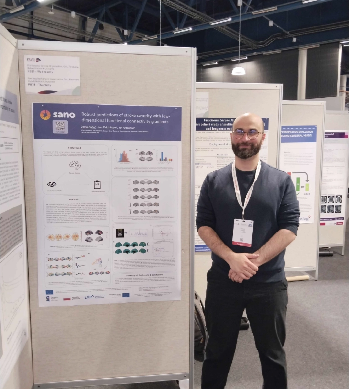
From May 21–23, 2025, the 11th European Stroke Organisation Conference (ESOC) took place in Helsinki — one of the most important scientific events dedicated to stroke research and treatment. Dr. Cemal Koba, Postdoctoral Researcher in Computational Neuroscience at Sano – Centre for Computational Medicine, presented a poster entitled:
“Robust Predictions of Stroke Severity with Low-Dimensional Functional Connectivity.”
Rethinking fMRI Use in Stroke Research
Functional MRI (fMRI) holds great potential for exploring brain dynamics after stroke, but its clinical applications remain limited. The challenges stem from the high dimensionality of fMRI data and the effects of disrupted cerebral blood flow. Dr. Koba and his co-authors proposed an innovative approach that addresses both of these challenges.
Methods: From Hemodynamic Lag to Functional Damage Mapping
The study involved 104 ischemic stroke patients and 26 healthy controls, all of whom underwent fMRI scanning within two weeks of stroke onset. After preprocessing, the data were adjusted for hemodynamic delays by shifting voxel-wise time series to align more closely with the gray matter signal.
Using nonlinear diffusion embedding, the researchers generated low-dimensional functional connectivity gradients. The first three components from the control group were averaged to define a reference topography. For each patient, damage was calculated as the Euclidean distance between their cortical connectivity profile and this reference. These damage maps were then used to predict stroke severity (NIHSS score). Gray matter disconnection profiles served as an additional reference.
Results: Accurate Prediction of Clinical Symptoms
Correcting for hemodynamic lag significantly enhanced functional connectivity, particularly along the visual–somatomotor axis (t(103)= 4.96, p<0.05). The derived maps of functional damage were strongly associated with specific clinical deficits, including language impairments (r=0.47), spatial neglect (r=0.41), and left-sided motor deficits (leg: r=0.49, arm: r=0.45). All associations were statistically significant after multiple comparison correction (pFDR<0.05).
Conclusion: Functional Damage Mapping as a Clinical Tool
The poster presented by Dr. Koba demonstrates that combining hemodynamic lag correction with dimensionality reduction techniques can improve the clinical relevance of fMRI in stroke. The resulting functional damage maps offer interpretable and anatomically accurate representations of stroke-induced brain changes — helping to bridge the gap between neuroimaging and clinical assessment.