Rosmary Blanco, Cemal Koba, Alessandro Crimi
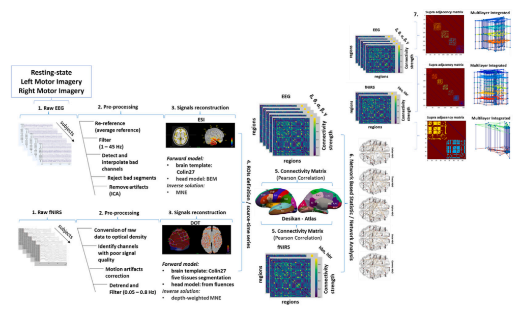
Rosmary Blanco, Cemal Koba, Alessandro Crimi Exploring the brain’s complex networks requires multiple neuroimaging techniques, each offering unique insights. Combining electroencephalography (EEG) and functional near-infrared spectroscopy (fNIRS) has gained attention for its potential to deepen our understanding of brain functioning. However, how these modalities relate is still an open question. Understanding how the electrical and hemodynamic activities relate is crucial for effectively integrating these modalities, potentially enhancing the spatio-temporal resolution of neuroimaging and revealing information about brain function that might be missed when each modality is used in isolation. In this study, we compared brain networks captured by EEG (electrical activity) and fNIRS (hemodynamic activity) in both resting and task-related conditions. Complementarity between modalities was observed, particularly during tasks, as well as a certain level of redundancy when comparing the multimodal and the unimodal approach, which depends on the modality and the specific brain state. Overall, the results highlight differences in how EEG and fNIRS capture brain network topology in different brain states and emphasize the value of integrating multiple modalities for a comprehensive view of brain functioning.
Authors: Rosmary Blanco, Cemal Koba, Alessandro Crimi
DOI: https://doi.org/10.1016/j.jocs.2024.102416
Link to article: www.sciencedirect.com
Keywords: multimodal neuroimaging, EEG, fNIRS, Multilayer Networks
READ HERE