Crisp edges, less noise: rethinking how CT images are softened.
Two new papers from the Computational Neuroscience team.
Every CT scan hides a trade-off that most patients, and even many researchers outside medical imaging, never hear about. Before a CT image reaches a radiologist, the scanner’s measurements must be transformed into a visual image using a mathematical reconstruction kernel. “Sharp” kernels produce crisp edges and reveal fine anatomical details, but they also amplify image noise. “Soft” kernels suppress noise, making images easier to interpret, but often at the expense of subtle structures.
Mahmoud Nasr and Jan K. Argasiński from Sano’s Computational Neuroscience team, together with Krzysztof Brzostowski and Adam Piórkowski, explored whether it is possible to achieve the best of both worlds: transforming sharp-kernel CT images into smoother, soft-kernel-like images after acquisition, without sacrificing diagnostically important anatomy and without requiring access to proprietary scanner raw data.
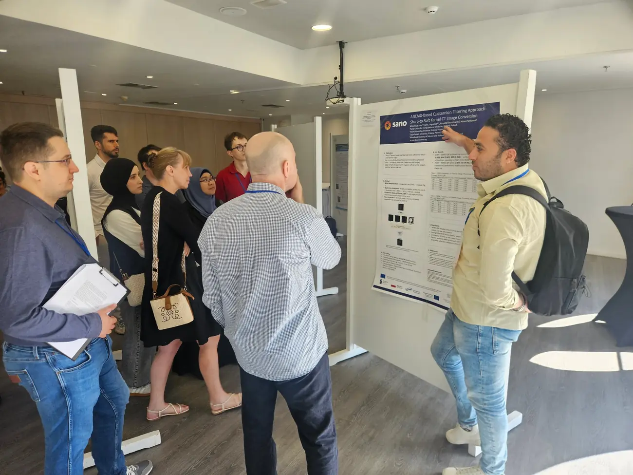
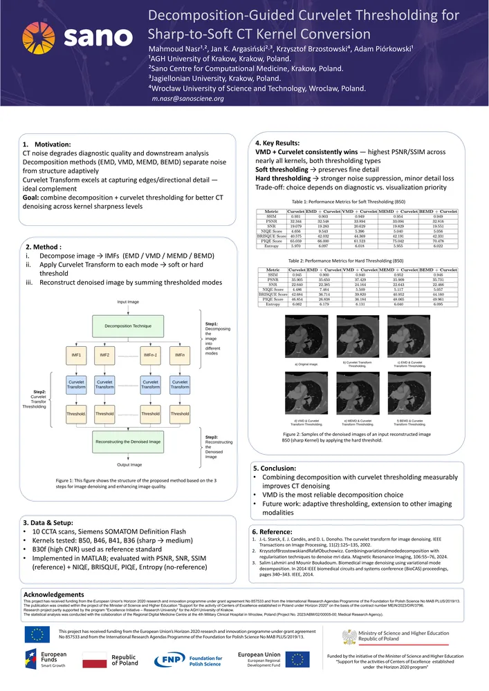
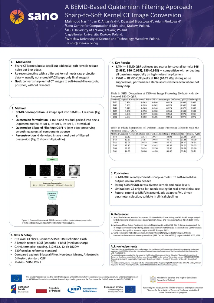
Their two papers, A BEMD-Based Quaternion Filtering Approach Sharp-to-Soft Kernel CT Image Conversion and Decomposition-Guided Curvelet Thresholding for Sharp-to-Soft CT Kernel Conversion, presented at ICCCI 2026 and published by Springer Nature, tackle this challenge using a common strategy: first decomposing an image into multiple layers representing different levels of detail, and then processing each layer according to its characteristics.
In the first study, the researchers combined Bidimensional Empirical Mode Decomposition (BEMD) with Quaternion Bilateral Filtering, a technique that allows information from different image components to be processed within a unified mathematical framework. Tested on CT images reconstructed using six different kernels, the method preserved structural information more effectively than several established denoising approaches, including Non-Local Means filtering, while achieving competitive noise reduction. Importantly, all of this was accomplished using only the reconstructed CT images themselves, with no need for access to the scanner’s original acquisition data.
The second study expanded the investigation by comparing four different decomposition techniques and combining them with curvelet thresholding, a method particularly well suited for preserving edges and curved anatomical structures while removing noise. Among the evaluated approaches, Variational Mode Decomposition (VMD) achieved the strongest performance based on standard image-quality metrics. The work also highlighted a practical trade-off relevant to future clinical applications: soft thresholding better preserves delicate image details, whereas hard thresholding provides stronger noise suppression.
Together, these two publications demonstrate the potential of decomposition-based image processing to improve CT image quality after reconstruction. By reducing noise while preserving anatomical detail, such techniques could support more consistent image analysis across clinical studies, facilitate retrospective research on existing datasets, and contribute to the development of more robust AI tools for medical imaging.
The presentation of two closely related studies at ICCCI 2026 highlights Sano’s ongoing contribution to advancing medical image processing methods at the intersection of computational intelligence and healthcare.
Here newsletter for the conference:
https://wit.pwr.edu.pl