A new paper by members of the Computational Neuroscience team at Sano, published in
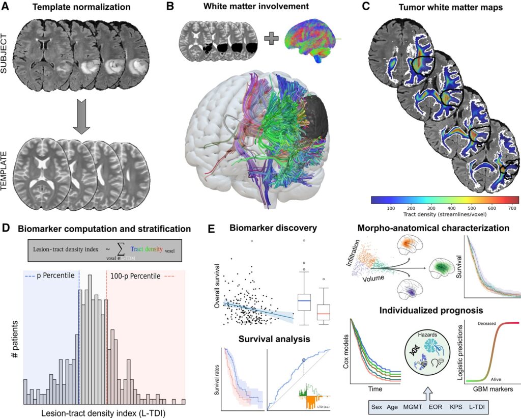
Neuro-Oncology, introduces a novel neuroimaging biomarker for glioblastoma (GBM) called
the Lesion-Tract Density Index (L-TDI). Moving beyond viewing GBM as a focal lesion, this
study treats it as a network disease that interacts with the brain’s white matter scaffold. By
analyzing large-scale white matter pathways in two independent patient cohorts, the
researchers found that L-TDI robustly stratifies survival rates and predicts outcomes more
accurately than traditional measures like tumor volume. This work marks a significant step
toward connectomics-guided neuro-oncology and improved individualized patient care.

The lesion-tract density index (L -TDI): a survival marker in human GBM. (A) For every subject, the data were normalized to a common template. The T2-weighted image was chosen as the reference modality, and the GBM was inversely masked to discard distorted tissue during the optimization. (B) The normalized tumor mask is used in combination with the whole brain normative tractogram to extract the set of streamlines that intersect the tumor in the same template space as in (A). (C) The lesion-tract density map (L -TDM) provides a unique white matter density map describing the tracts in (B) that interact with the GBM. (D) From the L -TDM, we can define the L -TDI marker by averaging the tract density within the L -TDM binary mask in (C). For a given cohort, the sample can be stratified according to a given percentile. (E) Once a stratification threshold has been set, we conduct thorough tests to assess the discovery of the biomarker. Additional survival analyses on multiple stratification thresholds help determine whether the potential biomarker holds prognostic value. The L -TDI can also be used to characterize both morphological and anatomical landscapes in GBMs, which can be used to understand differences in the survival rates. Once a robust effect has been identified, the L -TDI can be incorporated into standard risk analysis and predictive frameworks to improve patient care.
Image source: Figure 1 from “A non-local diffusion magnetic resonance imaging tract density index to capture network-level tumor–brain interactions”, Neuro-Oncology, Oxford University Press academic.oup.com/view-large/figure/556903333/noaf234f1.tif


See the full text:

Read the article

Autors: Joan Falcó-Roget, Gianpaolo Antonio Basile, Anna Janus, Sara Lillo, Letterio S Politi, Jan K Argasinski, Alberto Cacciola

Keywords: glioblastoma, brain connectome, diffusion tractography, glioblastoma, survival prediction