Karol Pustelnik
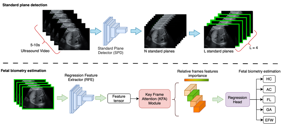
Fetal biometry assessment is essential for monitoring fetal growth and well-being. This is, however, challenging due to fetal move- ment and the arbitrary position of the fetus. Therefore, sonographer expertise is important to capture proper standard plane views of specific fetal body parts. Moreover, manual fetal biometry depends on operator skill and experience, which can cause interobserver variability and im- pact clinical decisions. Automatic fetal biometry may reduce the need for expertise to obtain accurate and reproducible measurements. We present a novel two-stage method for automated fetal ultrasound biometry us- ing a novel Key-Frame Attention framework (KFANet). Our proposed method automatically detects standard plane views and estimates fetal biometry measurements directly from 2D ultrasound videos. We evalu- ate our method using a clinical dataset of 2100 ultrasound videos from 700 patients and an external prospective dataset of 300 videos from 100 patients. We compare our deep learning-based estimations with man- ual measurements performed by six clinical experts and compare perfor- mance with other deep learning methods. Experimental results show that KFANet outperforms several state-of-the-art methods and estimates fetal biometry measurements with an accuracy comparable to clinical experts. The method has the potential to reduce the workload of sonographers and improve the accuracy and efficiency of fetal biometry assessment.

Fig1. The overview of KFANet for direct fetal biometry measurement estimation from ultrasound (US) video scans. In the first stage, the proposed method involves a Standard Plane Detector with a Spatial Attention Module to select standard planes of the fetal head, abdomen, and femur from US video scans. In the second stage, a Regressor Feature Extractor with a novel Key-Frame Attention module is applied to L selected standard planes to provide measurements of fetal body parts. On the output of KFANet, we obtain full fetal biometry measurements of head circumference (HC), abdominal circumference (AC), femur length (FL), gestational age (GA), and estimated fetal weight (EFW) for each patient. We compute EFW using Hadlock III formula.

