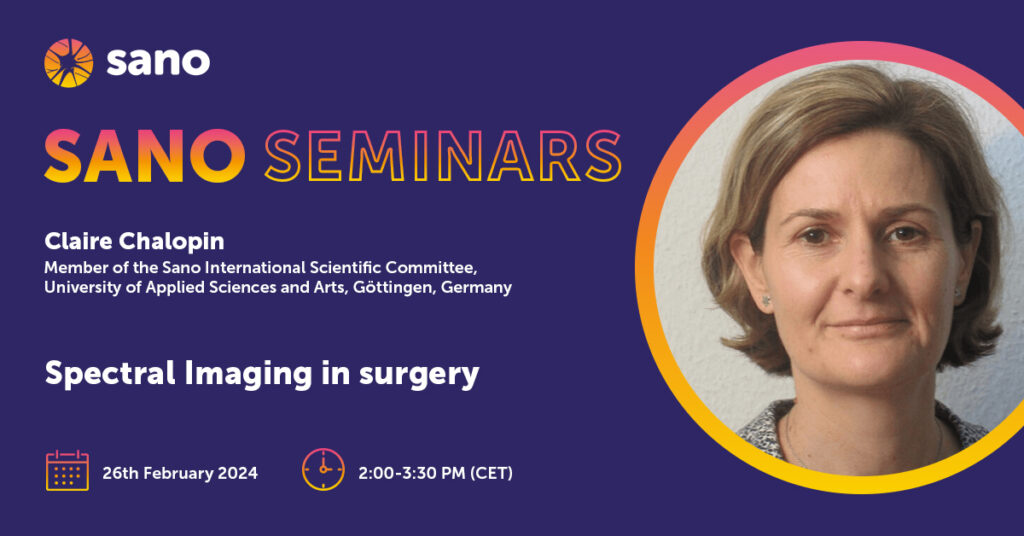
123. Spectral Imaging in surgery
Claire Chalopin – Member of the Sano International Scientific Committee, University of Applied Sciences and Arts, Göttingen, Germany
Abstract
Spectral imaging is a non-destructive imaging technique commonly used in various areas such as waste sorting and recycling, food quality and safety, pharmaceutics, agriculture and vegetation as well as environnmental monitoring. It was introduced in medicine 10 years ago in the context of research projects. The main medical applications are the estimation of tissue perfusion, the classification of tissue and the identification of tumor margins. The visualization of the information included in the spectral image data requires image processing techniques. Machine learning approaches are especially suitable due to the complexity of the data. Although medical spectral imaging showed very promising results, further studies are needed to acknowlede the first results, technically improve the devices and evaluate these new developments on patients.
In this presentation the following aspects will be developed:
- What are the physical and technical principles of spectral imaging? What are the advantages for the medicine and surgery?
- How are the spectral image data presented to physicians? Which algorithms are being developed for this purpose?
- What are the medical applications of spectral imaging?
- Which further medical and technical innovations are expected in the future?
About the author
Claire Chalopin studied physics at the University of Burgundy in France. She obtained her PhD degree at the University of Lyon in France in the field of medical image processing and analysis. Afterwards, she carried out a postdoc position at the Max-Planck-Institute for Human Cognitive and Brain Science of Leipzig in Germany, where she developed segmentation methods for medical image data. Then, she joined the Innovation Center for Computer Assisted Surgery of the Medical Faculty at the University of Leipzig. There she developed her own research topic and group for the development of multimodal imaging for surgery. She focussed on the clinical evaluation of new non-invasive imaging modalities and the development of innovative image analysis and visualization methods, especially using Artificial Intelligence approaches. In March 2023 she obtained a professor position in “data-driven imaging in medicine” at the University of Applied Sciences and Arts of Göttingen in Germany. She teaches students in medical engineering and continues her research in medical non-invasive imaging. Since October 2019 Claire Chalopin is a Member of the Sano International Scientific Committee.


